15 August, 2022 | 01:42 AM
https://www.dailynews.express/posts/in-monkeys-spike-based-vaccines-produce-some-immunity-based-on-cd8-t-cells
In monkeys, spike-based vaccines produce some immunity based on CD8 T-cells
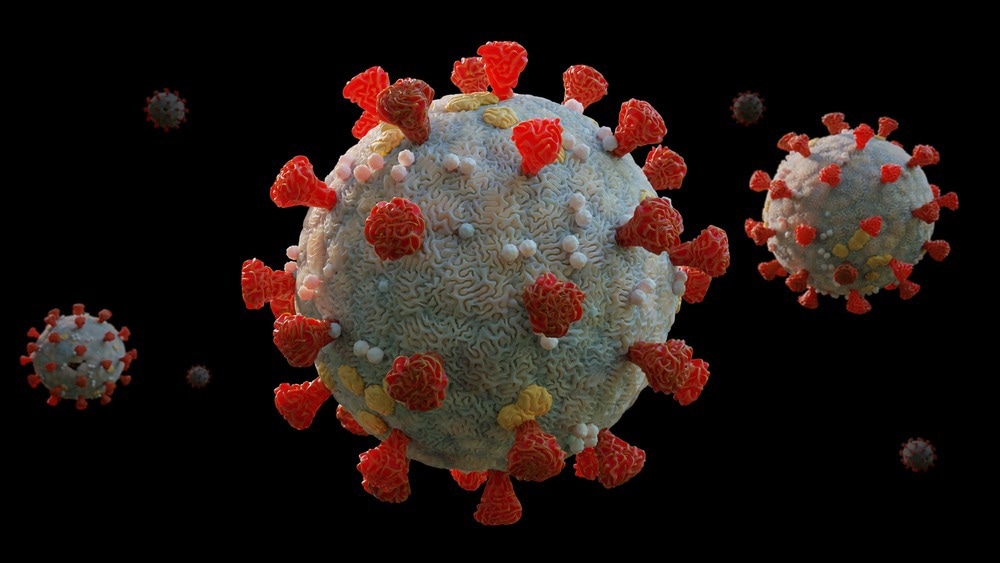
Scientists throughout the world are continually working to develop effective coronavirus disease 2019 (COVID-19) vaccines and therapeutics. The emergence of the severe acute respiratory syndrome coronavirus 2 (SARS-CoV-2) variants, such as the Omicron and Delta variants, have significantly reduced the efficacy of available vaccines. Study: CD8 T Cells Contribute to Vaccine Protection Against SARS-CoV-2 in Macaques. Image Credit: Dotted Yeti / Shutterstock.com The effectiveness of COVID-19 vaccines is often determined by the level of antibody responses elicited following vaccination. Preclinical and clinical studies have suggested that CD8+ T-cell responses are also associated with natural immune protection against SARS-CoV-2, especially when antibodies provide only partial protection. Greater durability and cross-reactivity have been reported for cellular immune responses as compared to neutralizing antibody (nAb) responses against SARS-CoV-2 variants. Importantly, both messenger ribonucleic acid (mRNA) and adenovirus vector-based vaccines are associated with 70% and 85% efficacy, respectively, against the Omicron BA.1 variant in the absence of Omicron-specific nAbs. Thus, other immune responses, aside from nAbs, have an important role in providing protection against severe COVID-19. Although virus-specific CD8+ T-cells can detect and remove infected cells, their direct function in vaccine protection against COVID-19 has not yet been determined. To date, all Phase III clinical trials of COVID-19 vaccines have excluded the evaluation of cellular immune responses as an immune correlate. In a recent Science Immunology study, scientists evaluate the role of CD8+ T-cells in vaccine protection against SARS-CoV-2 infection in rhesus macaques. A total of 30 adult male and female Rhesus macaques were allocated to six experimental groups. All animals were immunized with 5x10 10 viral particles of the Johnson & Johnson Ad26.COV2.S adenovirus vector-based vaccine, which is equivalent to a human dose of the vaccine. All test rhesus macaques were immunized through the intramuscular route at week zero. The test animals were subsequently injected with CD8-depleting monoclonal antibodies (mAbs) at week five, which was followed by the Delta variant challenge at week six. Each group received 50 mg/kg of the anti-CD8β CDR-grafted rhesus immunoglobulin G1 (IgG1) antibody (CD8b255R1), the anti-CD8α CDR-grafted rhesus IgG1 antibody (MT807R1), or an isotype-matched control antibody. Vaccination with Ad26.COV2.S elicited CD8+ T-cells that significantly contributed to controlling SARS-CoV-2 in a high dose heterologous challenge with the Delta variant in rhesus macaques. Immune responses following vaccination. Antibody responses at weeks 0, 4, and 6 following vaccination with Ad26.COV2.S and following challenge. A, Neutralizing antibody (NAb) titers by a luciferase-based pseudovirus neutralization assay. B , Receptor binding domain (RBD)-specific binding antibody titers by ELISA. C , Pooled peptide Spike-specific IFN-γ CD8 + and CD4 + T cell responses by intracellular cytokine staining assays at week 2 following vaccination with Ad26.COV2.S. Responses were measured against the SARS-CoV-2 WA1/2020 (black), B.1.617.2 (Delta; blue), and B.1.1.529 (Omicron; green) variants. Dotted lines represent limits of quantitation. Medians (red bars) are shown. In vaccinated animals, a reduction in CD8+ T-cells caused a higher viral load in the upper and lower respiratory tracts after animals were inoculated with the Delta variant. CD8α depletion had a greater impact on viral loads, perhaps because of the functional role of natural killer (NK) cells or CD8 depletion with the anti-CD8α mAb. Previous observations that BNT162b2 and Ad26.COV2.S vaccines provided significant protection against severe infection with Omicron BA.1 variant, in the absence of Omicron-specific nAbs, were similarly reported in the current study. Previous studies have also indicated that, unlike nAb responses, T-cell responses exhibit higher cross-reactivity against SARS-CoV-2 variants, including Omicron BA.1, which was also supported in the current study. Thus, these findings establish a definitive immunogenic context for clinical observations. Ad26.COV2.S vaccination induced CD8+ T-cell responses and contributed to controlling the viral load in rhesus macaques challenged with SARS-CoV-2 in a high-dose heterologous challenge model. The current model only focused on the virologic control in animals challenged with the SARS-CoV-2 Delta variant; thus, this animal model cannot be used to determine the role of CD8+ T-cell responses in providing protection during COVID-19. A previous macaque model-based study reported that higher antibody titers can prevent SARS-CoV-2 infection. However, all currently available COVID-19 vaccines show modest and brief efficacy in protecting individuals from contracting the SARS-CoV-2 Omicron variant, even after booster vaccination. In summary, the present study highlighted the significant contribution of CD8+ T-cell responses following vaccination with Ad26.COV2.S in providing protection against SARS- CoV-2 replication using a rhesus macaques model. The authors speculated that CD8+ T-cell responses also controlled the viral load after mRNA vaccination; however, this observation requires further experimental validation. Importantly, CD8+ T-cell responses adeptly restrict SARS-CoV-2 variants, such as the Delta and Omicron strains, which have been found to partially evade nAb responses. In the future, researchers must determine if CD8+ T-cell responses also positively affect SARS-CoV-2 vaccine protection in humans. Thus, further studies should also focus on T-cell responses, along with antibody titers, to evaluate vaccine efficacy in humans.